NA. V.I.KULAKOV»
Research Center for Obstetrics, Gynecology
and Perinatology named after Academician V.I.Kulakov»
Ministry of Health of the Russian Federation


FSBI «National Medical
Research Center for Obstetrics, Gynecology
and Perinatology named after Academician V.I.Kulakov»
Ministry of Health of the Russian Federation
Department of Oncopathology
Department of Oncopathology
Head of the Department: Vlada Kometova, Ph.D., E-mail: v_kometova@oparina4.ru
This is a multifunctional department, where the diagnosis of tumor and precancerous pathological changes in human cells and tissues is carried out.
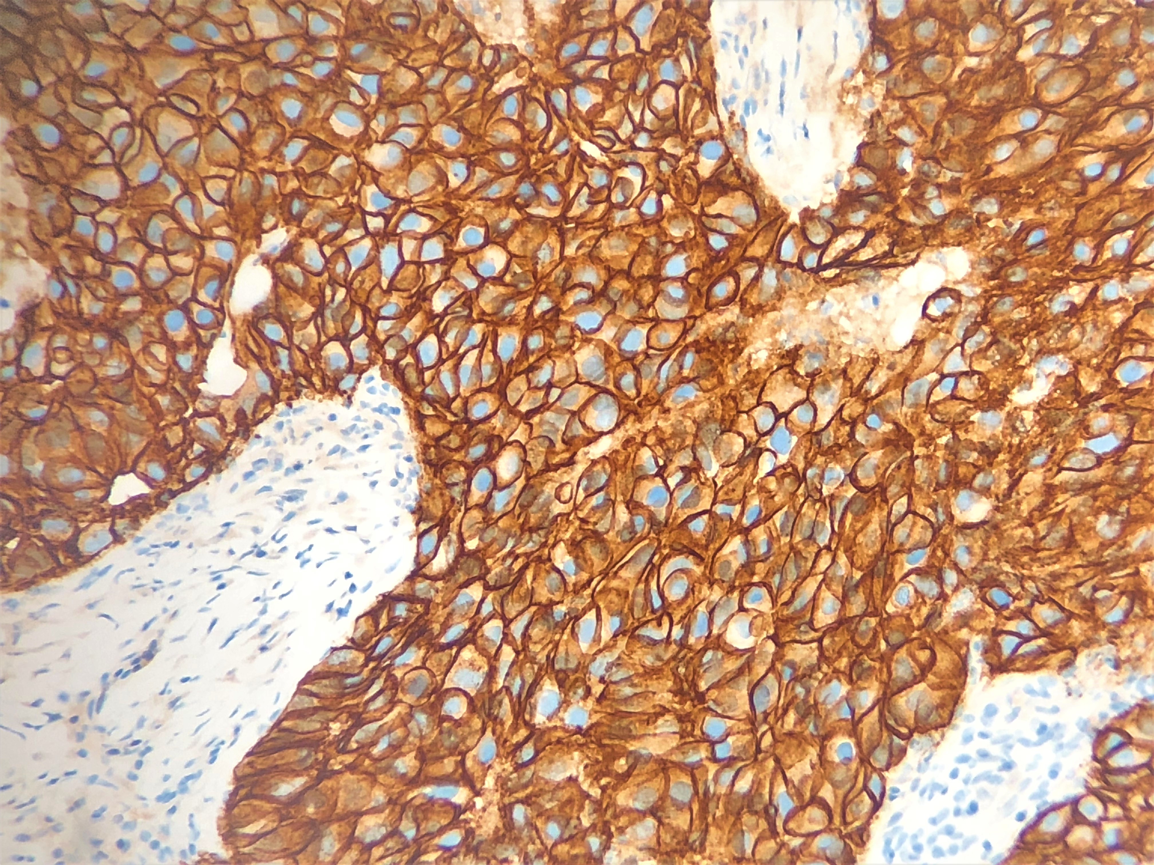
The Department provides a complex morphological and immunohistochemical analysis of bioptates and material obtained during oncological surgery, as well as autopsy material from patients with oncological diseases.
CLINICAL ACTIVITY
The experts of the Department carry out cytological and histological diagnostics of tumors and precancerous diseases of the female reproductive system:
- mammary glands;
- endometrium;
- myometrium;
- ovaries;
- cervix;
- fallopian tubes;
- vulva;
- vagina;
- pelvic peritoneum
FOCUS AREA
The Department of Oncopathology conducts research on “Tumor and pregnancy”, “Oncological pathology of uterus and gynecological endocrinology”, “BRСA-associated breast cancer and ovarian cancer”, “Lynch syndrome” “ART and tumors of the female reproductive system”, ”trophoblastic tumors”.
The Department of Oncopathology has a qualified medical and laboratory staff having extensive experience in diagnosing oncological diseases as well as in rare nosological forms and pathomorphological cases of gynecological oncological profile: immature teratomas and other ovarian germ cell tumors, especially benign and malignant tumors in pregnant women, juvenile fibroadenomas, gigantomastia, cancer and sarcoma in young women; PEComa, STUMP, specific variants of leiomyomas and other rare oncopathologies.
SCIENTIFIC WORK
Scientific activity includes:
- study of predictors of regional and distant metastasis of tumors of the female reproductive system;
- development of prognostic morphological and molecular genetic signatures of tumors of the female reproductive system;
- search for risk markers for development of breast, endometrial, cervical and ovarian cancer, which could be used for prediction of the disease;
- study of criteria for therapeutic pathomorphosis of tumors of the female reproductive system
- biobanking of tumor tissues of the female reproductive system for scientific research.
EQUIPMENT
The Department has modern equipment of leading world manufacturers: MacroPATH Pro-x digital imaging system (Milestone, Italy ), microtomes Leica (Germany) and Prestochill (Milestone, Italy), Microwave hybrid tissue processor LOGOS (Milestone, Италия), histological tissue processor “ASP 6025” (Leica Biosystems, Germany) and microtome ThermoScientific (Германия), research microscopes Olympus (Japan), Leica, Carl Zeiss (Germanyя, Nikon (Japan), «BD PrepStain» (USA) and «CellPrepPlus (Republik of kOREA» for preparation and staining of cervical cytology samples (Biodyne, Republicof Korea), staining system Ventana BenchMark ULTRA (ROSH, USA), Bond-III (Leica Biosystems, Germany) to detect atypical cells, digital slide scanner Aperio AT2.
This equipment and many other latest laboratory devices are used in the Department for cytological diagnostics of tumor and non-tumor pathologies, video and photo archiving of complicated cases with digital MacroPath D); presentation of scanned miscrocopic preparations during teleconcultations and video conferences; immunocytochemistry, biobanking of tissue for research work; immunohistochemical studies (there is a big set of primary monoclonal antibodies allowing to set any panel depending on the aim of the study); molecular and genetic analysis (SISH, FISH) and other studies in collaboration with other laboratories of the Center.